
Eye Anatomy
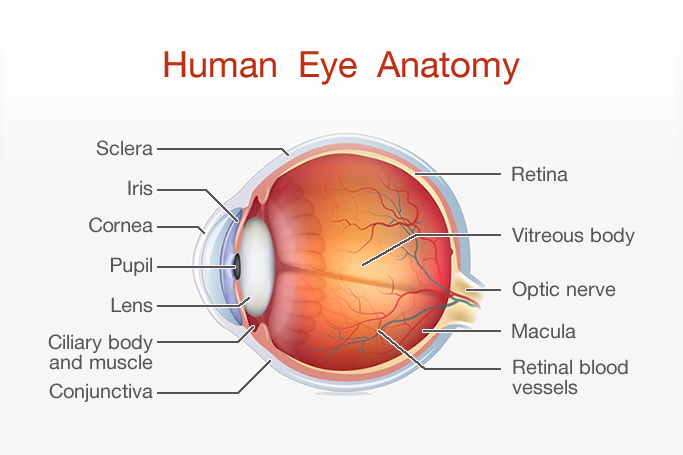
Human eye is like a camera, which needs a lens and a film to produce an image. Identically, the eyeball is using a lens (cornea, crystalline lens) to refract and focus the light, and a film (retina) on which to focus the rays. Focused rays are then converted into electrical impulse, transmitted through the optic nerve to the brain.
If one or several of the following major eye components are not functioning correctly, the result is a poor picture.
Each of them has a very specific role in the process of refraction:

Cornea
The cornea is the transparent, dome-shaped window covering the front of the eye. It is a powerful refracting surface. Like the crystal on a watch, it gives a clear window to look through. Because there are no blood vessels in the cornea, it is normally clear and has a shiny surface. The cornea is extremely sensitive - there are more nerve endings in the cornea than anywhere else in the human body.

Iris
The colored part of the eye is called the iris. It controls light levels inside the eye similar to the aperture on a camera. The round opening in the center of the iris is called the pupil. The iris is embedded with tiny muscles that dilate (widen) and constrict (narrow) the pupil size.

Pupil
The pupil is the opening in the center of the iris. The size of the pupil determines the amount of light that enters the eye. The pupil size is controlled by the dilator and sphincter muscles of the iris. Doctors often evaluate the reaction of pupils to light to determine a neurological function.

Lens
The crystalline lens is located just behind the iris. Its purpose is to focus light onto the retina. The nucleus, the innermost part of the lens, is surrounded by softer material called the cortex. The lens is encased in a capsular-like bag and suspended within the eye by tiny "guy wires" called zonules.

Retina
The retina is a multi-layered sensory tissue that lines the back of the eye. It contains millions of photoreceptors that capture light rays and convert them into electrical impulses. These impulses travel along the optic nerve to the brain where they are turned into images. There are two types of photoreceptors in the retina: rods and cones. The retina contains approximately 6 million cones. The cones are contained in the macula, the portion of the retina responsible for central vision. They are most densely packed within the fovea, the very center portion of the macula. Cones function best in bright light and allow us to appreciate color.

Optic Nerve
The optic nerve transmits electrical impulses from the retina to the brain. It connects to the back of the eye near the macula. When examining the back of the eye, a portion of the optic nerve called the optic disc can be seen
Rutnin-Gimbel LASIK Centre integrates over 60 years of expertise in eye care from the profressionals of Rutnin Eye Hospital
Contact Details
Quick Links
Working Hours


66 Nuam Building Fl. 16
Sukhumvit 21 (Asoke)Rd., Khlongtoey Nua, Wattana, Bangkok 10110
66 Nuam Building Fl. 16
Sukhumvit 21 (Asoke)Rd., Khlongtoey Nua, Wattana, Bangkok 10110